Pneumonia is an infection that can cause inflammation and fluid buildup in one or both lungs. It is a common illness, particularly among the elderly and those with weakened immune systems. Diagnosing pneumonia can be challenging, as symptoms may overlap with other respiratory illnesses.
What is a Chest X-Ray?
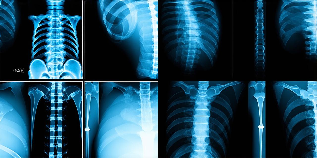
A chest X-ray is an imaging test that uses electromagnetic radiation to create a picture of the inside of the chest cavity. It can help doctors identify any abnormalities or lesions in the lungs, such as tumors, pneumonia, or fluid buildup. Chest X-rays are quick and easy to perform, making them a valuable tool for diagnosing respiratory illnesses.
How is a Chest X-Ray Performed?
During a chest X-ray, the patient will be asked to lie down on a table with their arms extended above their head. The radiologist will then position the X-ray machine over the chest, and a beam of radiation will pass through the body. This radiation is absorbed differently by different tissues in the body, creating an image on a special type of film. The radiologist will then interpret the image to determine if there are any abnormalities present.
Common Causes of Pneumonia
Pneumonia can be caused by a variety of factors, including viral infections, bacterial infections, and certain chemicals or particles in the air. The most common causes of pneumonia include influenza, Streptococcus pneumoniae, and Mycoplasma pneumoniae.
Symptoms of Pneumonia
Pneumonia can cause a wide range of symptoms, including coughing, shortness of breath, chest pain, fever, and fatigue. In severe cases, it may also cause fluid buildup in the lungs, leading to difficulty breathing and even life-threatening complications.
Diagnosis of Pneumonia
The diagnosis of pneumonia is typically based on a combination of symptoms, medical history, and laboratory tests. Chest X-rays are often used to help diagnose pneumonia by identifying any fluid buildup or abnormalities in the lungs. In some cases, additional imaging tests, such as CT scans or ultrasounds, may also be used to further evaluate the lungs.
Treatment of Pneumonia
The treatment of pneumonia typically involves a combination of antibiotics, fluids, and oxygen therapy to help the patient breathe more easily. In severe cases, hospitalization may be necessary to provide close monitoring and intravenous (IV) fluids and medications.
Conclusion
Chest X-rays are an important tool for diagnosing pneumonia and monitoring its progression. They can help doctors identify any abnormalities or fluid buildup in the lungs, allowing for early intervention and better treatment outcomes. Pneumonia is a common respiratory illness that can be caused by a variety of factors. Symptoms may include coughing, shortness of breath, chest pain, fever, and fatigue. Diagnosis is typically based on a combination of symptoms, medical history, and laboratory tests, including chest X-rays. Treatment typically involves antibiotics, fluids, and oxygen therapy, with hospitalization possible in severe cases.